ERTH3104 Virtual Field Trip to New England Orogen
Stop name: Chaffey Dam
Location: The Chaffey Dam along the Peel River; outcrops along the Nundle Road. Link to the Chaffey Dam project from the NSW Government website.
Coordinates: 31°20'48.11"S, 151° 8'33.76"E
What to see: A beautiful OPS section is exposed here along the road cut. This OPS section contains slices of rocks of varying lithologies, including basalt, clastic sediment, and red chert. Structural characteristics of OPS are best seen at this location. Slices of ocean floor rocks are now in faulted contact with each other and thickness of rock slices could be as thin as < 1 m. Consider the formation mechanism of OPS and discuss why these rocks are now closely faulted together.

(Above) OPS outcrop at Chaffey Dam (Photo by J. Aitchison) Click here to enter 360 view.
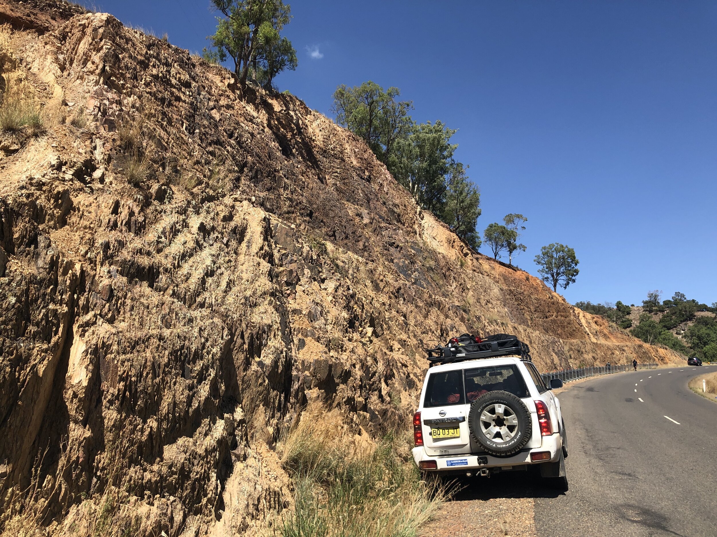
(Above) An overview of the road cut. Note that the OPS slices are all near vertical with near parallel bedding and foliation. (Photo by R. Zhou)

(Above) A close-up look a this outcrop. The green layer is an altered basalt. The red layer (right to the green layer) is a red chert. (Photo by R. Zhou)

(Above) A deeply weathered layer of rocks (a fine-grained mafic igneous rock) highlighting the difference in rock types within OPS. (Photo by R. Zhou)

(Above) Intensely deformed chert. (Photo by Y. Chen)

(Above) Isoclinal folds in chert. (Photo by Y. Chen)

Dating OPS: The most commonly used method to date the OPS is micropaleontology (biostratigraphy). Radiolarians are silica-secreting, single-celled protists that dwell in open-ocean locations. They occur throughout the water column from near surface to great depths since the Paleozoic. Radiolarians could be extracted from marine facies rocks, including
red ribben chert, limestone and even mudstone, of which chert is the most common rock type.
Photo to the left shows several examples of modern radiolarians. Each of them is about 100-200 micrometres in size! (Photo by S. Kachovich)
(Above) 3D model of a radiolarian. With the modern imaging techniques such as micro-CT scanning, we are able to much better characterize the structure of radiolarians, particularly their internal strucures. This 3D model was made by J. Sheng.